1946 syntynyt Tampereella
Ylioppilastutkinto 1964 Lempäälä
Lääketietaan kandidaatti 1966 Turun yliopisto
Lääketieteen lisensiaatti 1972 Turun Yliopisto
Dietetiikan opiskelu 1998 - 2001 Göteborgin Yliopisto
Eläkkeelle 2010
J Neurosci. 2011 Jun 22;31(25):9404-13.Unesterified cholesterol accumulation in late endosomes/lysosomes causes neurodegeneration and is prevented by driving cholesterol export from this compartment.
Department of Pediatrics, The University of Texas Southwestern Medical School, Dallas, Texas 75390, USA. Abstract
While unesterified cholesterol (C) is essential for remodeling neuronal plasma membranes, its role in certain neurodegenerative disorders remains poorly defined. Uptake of sterol from pericellular fluid requires processing that involves two lysosomal proteins, lysosomal acid lipase, which hydrolyzes C esters, and NPC1 (Niemann-Pick type C1). In systemic tissues, inactivation of either protein led to sterol accumulation and cell death, but in the brain, inactivation of only NPC1 caused C sequestration and neurodegeneration. When injected into the CNS of the npc1(-/-) mouse, 2-hydroxypropyl-β-cyclodextrin (HP-β-CD), a compound known to prevent this C accumulation, diffused throughout the brain and was excreted with a t(½) of 6.5 h. This agent caused suppression of C synthesis, elevation of C esters, suppression of sterol regulatory-binding protein 2 (SREBP2) target genes, and activation of liver X receptor-controlled genes. These findings indicated that HP-β-CD promoted movement of the sequestered C from lysosomes to the metabolically active pool of C in the cytosolic compartment of cells in the CNS. The ED(50) for this agent in the brain was ∼0.5 mg/kg, and the therapeutic effect lasted >7 d. Continuous infusion of HP-β-CD into the ventricular system of npc1(-/-) animals between 3 and 7 weeks of age normalized the biochemical abnormalities and completely prevented the expected neurodegeneration. These studies support the concept that neurons continuously acquire C from interstitial fluid to permit plasma membrane turnover and remodeling. Inactivation of NPC1 leads to lysosomal C sequestration and neurodegeneration, but this is prevented by the continuous, direct administration of HP-β-CD into the CNS.
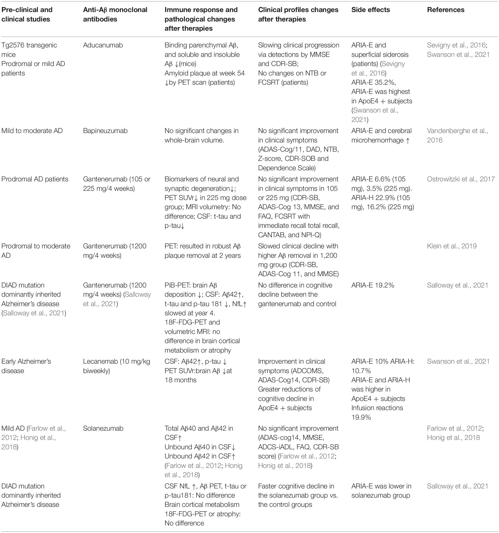
Swanson CJ, Zhang Y,
Dhadda S, Wang J, Kaplow J, Lai RYK, Lannfelt L, Bradley H, Rabe M,
Koyama A, Reyderman L, Berry DA, Berry S, Gordon R, Kramer LD, Cummings
JL.Alzheimers Res Ther. 2021 Apr 17;13(1):80. doi: 10.1186/s13195-021-00813-8.PMID: 33865446Free PMC article.Clinical Trial.
BACKGROUND: Lecanemab (BAN2401), an IgG1 monoclonal
antibody, preferentially targets soluble aggregated amyloid beta
(Abeta), with activity across oligomers, protofibrils, and insoluble
fibrils. ...CSF biomarkers were supportive of a treatment effect. Lecanemab was …
van Dyck CH, Swanson
CJ, Aisen P, Bateman RJ, Chen C, Gee M, Kanekiyo M, Li D, Reyderman L,
Cohen S, Froelich L, Katayama S, Sabbagh M, Vellas B, Watson D, Dhadda
S, Irizarry M, Kramer LD, Iwatsubo T.N Engl J Med. 2023 Jan 5;388(1):9-21. doi: 10.1056/NEJMoa2212948. Epub 2022 Nov 29.PMID: 36449413Clinical Trial.
Participants were randomly assigned in a 1:1 ratio to receive intravenous lecanemab (10 mg per kilogram of body weight every 2 weeks) or placebo. ...Lecanemab resulted in infusion-related reactions in 26.4% of the participants and amyloid-related imaging abnormaliti …
Lecanemab (lecanemab-irmb; LEQEMBI) is
a humanized immunoglobulin gamma 1 (IgG1) against aggregated soluble
and insoluble forms of amyloid-beta peptide. ...This article summarizes
the milestones in the development of lecanemab leading to this first approval f …
McDade E, Cummings JL, Dhadda S, Swanson CJ, Reyderman L, Kanekiyo M, Koyama A, Irizarry M, Kramer LD, Bateman RJ.Alzheimers Res Ther. 2022 Dec 21;14(1):191. doi: 10.1186/s13195-022-01124-2.PMID: 36544184Free PMC article.Clinical Trial.
The objective of this analysis is to report results from
study 201 blinded period (core), the open-label extension (OLE), and gap
period (between core and OLE) supporting the effectiveness of lecanemab. METHODS: The lecanemab study 201 core was a double-blind, rando …
Tahami Monfared AA, Tafazzoli A, Ye W, Chavan A, Zhang Q.Neurol Ther. 2022 Jun;11(2):863-880. doi: 10.1007/s40120-022-00350-y. Epub 2022 Apr 25.PMID: 35469060Free PMC article.
The mean time to mild, moderate, and severe AD dementia was longer for patients in the lecanemab
+ SoC group than for patients in the SoC group by 2.51, 3.13, and 2.34
years, respectively. ...The model also predicted a lower lifetime
probability of admission to institution …
All three antibodies bound monomers with low affinity. However, lecanemab and aducanumab had very weak binding to monomers, and gantenerumab somewhat stronger binding. Lecanemab was distinctive as it had tenfold stronger binding to protofibrils compared to fibrils. …
Ihmisen keho tarvitsee rakenteellisen aminotypen ja siihen kuuluvan aineenvaihdunnan lisäksi paljon funktionaalisia typpeä siältäviä ja käsitteleviä proteiineja. Tässä keskityn NO- molekyyliin , typpioksiiin, jota keho tarvitsee myös. ennenkin olen kirjoittanut tetrahydrobiopteriinista, joten tässä kertaan asiaa. Tgerahydrobiopteriini on smantapainen kuin foolihappo, muta eroaa siitä, että sitä pystyy keho valmistamaan itse, jos aineenvaihdunta on optimaali. Foolihapon saanti kuitenkin tukee sen muodostumsita ja funktiota. MAhdollsiesti se kuitenkin voi olla myös vitamiini joissain tilanteissa. Ainakin pitää tunnistaa, milloin tetrahydrobiopteriinin muodostuminen kehosa kompromittoituu ja siitä tulee funktionaalinen puutos.
Typpioksidisyntaasi (NOS) on kriittinen entsyymi välittäjäainemolekyylin NO muodostumisessa L-arginiiniaminohaposta. NOS-entsyymi vaatii kofaktoriksi tetrahydrobiopteriinia typpioksidimolekyylin muodostamiseksi. NO-syntaasi on on yksi harvoista entsyymeistä , joka käyttää juuri tätä kofaktoria . Sen lisäksi tetrahydrobiopteriinin merkitys NOS-entyymin katalyyttisessä mekanismissakin poikkeaa muiden entsyymien katalyyttisistä mekanismeista. NOS-entsyymin katalyyttisen syklin aikana tetrahydrobiopteriini muodostaa radikaalilajinsa, joka sitten taas redusoituu palauttaen sen tehokkaasti ennalleen jokaisen NO-synteesisyklin jälkeen.
Tässä katsauksessa tehdään yhteenvetoa siitä tiedosta, mitä meillä nyukyään on tetrahydrobiopteriinin roolista NOS-entsyymien rakenteessa, fuktiossa ja katalyyttisessä mekanismissa.
Nitric oxide synthase (NOS) is a critical enzyme for the
production of the messenger molecule nitric oxide (NO) from L-arginine.
NOS enzymes require tetrahydrobiopterin as a cofactor for NO synthesis.
Besides being one of the few enzymes to use this cofactor, the role of
tetrahydrobiopterin in NOS catalytic mechanism is different from other
enzymes: during the catalytic cycle of NOS, tetrahydrobiopterin forms a
radical species that is again reduced, thus effectively regenerating
after each NO synthesis cycle.
NO-syntaasit, typpioksidin syntetisoijat, ovat entsyymeitä, jotka katalysoivat typpioksidin NO muodsotusta arginiini (arg, R) -nimisestä aminohaposta . NO on tärkeä signaloiva molekyyli , joka osallistuu lukuisiin biologisiin prosesseihin kuten hermoimpulssien välittämiseen, verisuonten laajenemiseen ja immuunivasteeseen. NOS-entsyymiproteiinit ovat aktiiveja kun ne ovat homodimeerimuodossa. Yksi monomeeri käsittää kaksi hyvin selvästi määriteltävää domeenia. Toinen on N-terminaalinen ( aminoterminaali) alue. Se on oxygenaasidomeeni, sitoo hemiä ja L-arginiinia sekä tetrahydrobiopteriinia (H4B, toinen merkintä: BH4). Toinen domeeni on C-terminaalinen (karboksyyliterminaali) alue. Se on reduktaasidomeeni ja sitoo flaviiniadeniinidinukleotidia (FAD) flaviinimononukleotidia (FMN) ja nikotinamidiadeniinidinukleotidia (NADPH) ( jotka ovat B-vitamiineista kehossa muodostettuja koentsyymeitä), ja toimittaa elektoroneja NADPH:sta käsin oxygenaasidomeenilleen. Molemmat domeenit voivat ilmentyä erikseen ja ne ovat myös puhdistettavissa erikseen ja niiden ligandien sitomistapa ja reaktiivisuus on selvitetty. Oxygenaasi ja reduktaasidomeenien välissä on lyhyt sekvenssi (30-40 aminohapon jakso) ja se antaa sitoutumiskohdan kalmoduliiniproteiinille (CaM). KUVA allaolevassa linkissä Fig.1.
Nitric oxide synthases (NOS, EC 1.14.1.39) are enzymes that catalyze the formation of nitric oxide (NO) from L-arginine.
NO is an important signaling molecule that participates in a number of
biological processes, including neurotransmission, vasodilation, and
immune response (1).
NOS proteins are active as homodimers, and each monomer consists of two
well-defined domains. The N-terminal oxygenase domain binds heme, L-arginine, and (6R-)5,6,7,8-tetrahydrobiopterin (H4B).
The C-terminal reductase domain binds flavin adenine dinucleotide
(FAD), flavin mononucleotide (FMN), and nicotinamide adenine
dinucleotide phosphate (NADPH) and provides electrons to the oxygenase
domain from NADPH. Both domains can be expressed and purified
separately, and conserve their ligand binding and reactivity. Between
the oxygenase and reductase domains, a short sequence (30–40 aminoacids)
provides a binding site for calmodulin (CaM) (Fig. 1).
Schematic mechanism of electron transfer in NOS enzymes. In the presence of H4B,
the oxygenase domains (red ovals) of two NOS monomers are usually in a
dimeric form stabilized by the oxygenase (heme) domains. Top, in the
absence of CaM, there is limited electron transfer through the NOS
reductase domain (rectangles); NADPH reduces FAD via hydride transfer
and then electrons are transferred from FAD to FMN. The electron
transfer from FMN to the heme is impaired. Bottom, when CaM binds to the
NOS it helps stabilize a conformation where the electron transfer from
FMN to heme is enabled and the oxygenase domain can catalyze NO
synthesis from L-Arginine. [Color figure can be viewed in the online issue, which is available at wileyonlinelibrary.com.]
The oxygenase domain of NOS catalyzes the oxidation of L-arginine to L-citrulline and NO in two sequential oxidation steps (Scheme 1). In the first step, L-arginine is hydroxylated to make Nω-hydroxy-L-arginine (NOHA) in a process that requires one molecule of NADPH and one molecule of oxygen per mol of L-arginine reacted. In the second step, NOHA is oxidized to L-citrulline and NO and 1 molecule of oxygen and 0.5 molecules of NADPH are required.
NOS converts L-Arginine to L-Citrulline and NO in a two-step process.
NOS converts L-Arginine to L-Citrulline and NO in a two-step process.
The three mammalian isoforms of NOS show high sequence
homology but greatly differ in their localization and regulation. The
inducible isoform (iNOS) has a high affinity for CaM and is
constitutively active; this isoform is mainly regulated at the
transcriptional level. Endothelial (eNOS) and neuronal (nNOS) enzymes
are constitutively expressed and are reversibly activated by calcium
through the binding of Ca2+-containing CaM. Related proteins have been discovered in bacteria but consisting of only the oxygenase domain (2).
The bacterial NOS enzymes also contain heme but are less stringent in
their pterin requirements and can often bind tetrahydrofolate (H4F) and H4B
with similar affinity. Due to the absence of a connected reductase
domain, they must receive electrons from other electron transfer
proteins (2-4). The recently discovered NOS from Sorangium cellulosum is a notable exception to this rule (5).
The general biochemistry of NOS enzymes has been extensively reviewed from the biophysical (6-9) and clinical perspective (10). Reviews on H4B biochemistry have also treated NOS (11, 12). In this review, we will focus on the function of tetrahydrobiopterin in NOS enzymes.
H4B Requirement for NOS Catalysis
Early studies of NOS enzymes identified H4B as a necessary cofactor for NO synthesis (13-15) with a stoichiometry of one molecule of H4B per subunit (16). Nevertheless, the function of H4B was controversial, with a variety of suggested roles. A redox cycle involving H4B and H2B, as in aromatic amino acid hydroxylases was considered, but this would require one H4B molecule per cycle, in contradiction with experimental data (15, 17). Strong evidence indicated that H4B enhanced dimer formation (18, 19).
However, the evidences of some redox effect did slowly accumulate.
Single turnover experiments indicated that the stability of the heme-oxy
complex was dependent on H4B presence, and H4B was required for the reaction to advance past the formation of a ferrous dioxygen/ferric superoxide complex (20). Other results indicated that H4B analogs were able to catalyze L-arginine hydroxylation to a variable extent, but all were reduced forms (21). It was even unclear if H4B was required for the NOHA oxidation step (22). The detection of a H4B radical (23-26) and the coupling of this radical formation with product formation (26)
finally lead to the currently accepted notion—the decay of the ferrous
dioxygen/ferric superoxide complex is linked to the formation of a H4B radical species (8, 9, 27, 28).
H4B Binding in NOS
Pterin Binding Affinity of NOS
NOS enzymes bind H4B with high affinity. The presence of significant amounts of H4B in the enzyme after purification evinces the tight binding of the cofactor by the native protein (13-15). The binding of L-arginine and H4B shows a synergistic effect: prior binding of L-arginine increases the binding affinity for H4B and vice versa (21, 29, 30). Mammalian NOS enzymes bind H4B with affinities in the nM range. H4B is usually the preferred substrate over H2B, except for the eNOS enzyme (31). This fact has important implications for human pathology (10, 12, 31). Bacterial NOS enzymes operate in a wide range of KD values, and sometimes bind H4F with higher affinity (2, 32, 33). It should be noted that although many other pterins can bind to NOS (see (11) and references therein), only a few H4B analogs can support NO synthesis (21). The binding affinities for several pterins are shown in Table 1.
Table 1.
Dissociation constants for pterin binding to NOS enzymes
Pterin Binding Pocket in Mammalian and Bacterial NOS
Structures of the oxygenase domains for the three eukaryotic NOS isoforms (34-36) and several bacterial NOS proteins (37-39)
have been reported. The alignment of the sequences for these NOS
proteins indicates the notable similarity between mammalian and
bacterial NOS enzymes (Fig. 2).
The presence of additional elements in the N-termini of the mammalian
proteins can be also noted; these motifs are involved in dimer formation
and pterin binding (Figs. 2 and 3).
The available structures clearly show that the absence of this
N-termini allows bacterial NOS proteins to bind larger pterins (Fig. 3). The conservation of the H4B
binding environments in eNOS, iNOS, and nNOS is remarkable. Moreover,
the bacterial NOS proteins also share many similarities in the pterin
binding region. H4B is bound to NOS by an extensive network of hydrogen bonds, with a highly conserved pattern for the three NOS isoforms (34-36) (Fig. 3).
Most hydrogen bonds are contributed by the protein but there is also an
interaction with the heme group. Several hydrogen bonds are contributed
by the protein backbone and the side chain of a conserved Arg residue
(Arg375 in mouse iNOS). A conserved tryptophan residue also provides
stabilization through a π-stacking interaction (Trp457 in mouse iNOS).
Notably both Arg and Trp residues are conserved in mammalian and
bacterial NOS proteins. The pterin binding pocket is placed in the
oxygenase dimer interface. This location clearly suggest a relationship
with the role of H4B binding on stabilization of the NOS dimer.
Sequence alignment of mammalian and bacterial
NOS proteins. Regions involved in dimerization are shown in yellow;
Zn-binding cysteines are shown in green; residues directly binding H4B or H4F
are shown in red and blue to indicate that they belong to different
monomers. Note the additional N-terminus structure for the mammalian NOS
proteins, including the N-terminal hook region and the Znbinding
motif. nNOS, rat nNOS; iNOS, mouse iNOS eNOS, bovine eNOS; bsNOS, Bacillus subtilis NOS, saNOS, Staphylococcus aureus NOS; gsNOS, Geobacillus stearothermophilus NOS. [Color figure can be viewed in the online issue, which is available at wileyonlinelibrary.com.]
Protein environment of the H4B/H4F
cofactor. Panels A and B, comparison of the overall structures of iNOS
(panel A) and bsNOS (Panel B). Monomers are shown in blue and yellow.
The extra N-terminal portion of iNOS, not present in bacterial NOS, is
shown in red. Heme (pink), H4B (H4F for bsNOS, yellow) and L-Arg (NOHA for bsNOS, blue) are shown as sticks. Panels C and D, detail of the interactions of the H4B/H4F cofactor. Panel C, Several iNOS residues and the heme group form a hydrogen bonding network with the H4B cofactor. Relevant iNOS residues are shown in green and grey; H4B (yellow), Heme (pink), and the substrate L-Arginine
(blue) are shown as sticks. Trp455 and Phe470 are shown in grey color
to indicate that they belong to the other monomer. Two water molecules
that form H-bonding interactions with H4B are shown as red
spheres. The hydrogen bonding interactions are shown as yellow dashes.
Ser112, Ile456, and Trp457 make H-bonds through the main chain carbonyl
groups; Arg375 interacts through its side chain. The side chain of
Trp457 forms a π-stacking interaction with H4B. Panel D, Several bsNOS residues and the heme group form a hydrogen bonding network with the H4F cofactor. Relevant bsNOS residues are shown in green and grey; H4F
(yellow), Heme (pink), and the substrate NOHA (blue) are shown as
sticks. Trp323 and Phe338 are shown in grey color to indicate that they
belong to the other monomer. The hydrogen bonding interactions are shown
as yellow dashes. Thr324 and Trp325 make H-bonds through the main chain
carbonyl groups; Arg243 interacts through its side chain. The side
chain of Trp325 forms a π-stacking interaction with H4F. The figure was made using PyMOL (http://www.pymol.org/) and the crystal structure of the mouse iNOSoxy dimer (PDB entry 1NOD (34)) and the bsNOS dimer (PDB entry 1M7Z (38)). [Color figure can be viewed in the online issue, which is available at wileyonlinelibrary.com.]
H4B Function in NOS Catalysis
H4B Role in the L-Arginine Hydroxylation Step
Despite initial difficulties to assess the role of H4B in NOS catalysis, the study of the formation of NOHA from L-arginine yielded increasing evidence of a role for H4B beyond the H4B–H2B cycle of aromatic hydroxylases. The presence of H4B increases the rate of the FeIIO2 complex decay, a novel role for H4B (20). Other studies observed that NOHA formation only occurred in the presence of H4B (40, 41). As previously pointed out, the observation of a H4B radical formation during the reaction of NOS with L-arginine clearly indicated the active role of H4B during the formation of NOHA from L-arginine (23-26). The observation of the build-up of an H4B radical as the ferrous oxygen complex decays was instrumental in establishing a coherent mechanism (26) (Fig. 4).
Time course of the formation of the oxygen complex (FeIIO2), oxidized heme (FeIII), NOHA, and H4B radical during the single turnover of iNOS. Traces for FeIIO2 and FeIII are determined by stopped-flow experiments; NOHA is determined from rapid-quench experiments; H4B radical is determined by Electron Paramagnetic Resonance on rapid freeze experiments. Data from (26). [Color figure can be viewed in the online issue, which is available at wileyonlinelibrary.com.]
First steps of the reaction were clear, with the
reductase domain of NOS providing the electrons to reduce the heme, and
subsequent oxygen binding to form a ferrous-dioxygen complex (FeIIO2). As this species decays with concomitant formation of an H4B radical, a one electron transfer from H4B
to the heme-oxy complex with the formation of a ferric peroxo species
is expected. From this point, different reactions are possible. By
analogy with P450 systems, protonation of this species to form the
ferric-hydroperoxo species and eventual formation of the FeIVO porphyrin radical (Compound I) species was proposed (Fig. 5). In this process, the H4B radical is still present after L-arginine hydroxylation, and an additional electron—provided by the flavoprotein domain (42)—is needed to return to the initial resting state.
Putative mechanism for the hydroxylation of L-Arginine
by NOS. Boxes in continuous lines indicate stable species, boxes in
dotted lines indicate transient intermediate species. The oxidation
state of H4B and the substrate bound (L-Arginine/NOHA) are shown in the top left and right, respectively, of each species. The transfer of an electron from H4B
to the ferrous-oxy species triggers the formation of a ferric-peroxy
species that will capture two protons and eliminate a water molecule to
form a compound I-like species that will carry out the hydroxylation of
the substrate L-Arginine. Note that the H4B
radical (red) is not reduced after NOHA formation and has to receive
one electron from the flavin domain to continue the catalytic cycle.
[Color figure can be viewed in the online issue, which is available at
wileyonlinelibrary.com.
H4B Role in the NOHA Oxidation Step
The formation of citrulline and NO from NOHA has many similarities with the process of L-arginine oxidation to NOHA and also substantial differences. We must consider that the oxidation of L-arginine
to NOHA requires two electrons and two protons, but the reaction of
NOHA to citrulline and NO requires one electron, as a two electron
donation would lead to the formation of nitroxyl (NO−) species as final product. Therefore, it was considered that the electron transfer from H4B may not be necessary, and even detrimental, for this step.
Extensive evidence further indicates that H4B is necessary for NOHA oxidation (43-45). Interestingly, reaction of nNOS with NOHA and hydrogen peroxide in the absence of H4B leads to the formation of nitroxyl, whereas H4B directs the reaction toward NO instead (46). Therefore, it was proposed that the H4B radical formed in the first steps of the reaction could actually be reduced by a FeII–NO intermediate to produce the observed FeIII-NO species (24, 46, 47). Later reports are consistent with such model (44, 48-50).
These observations indicate that the requirement for H4B in the NOHA oxidation step appears to be two-pronged (Fig. 6). First, H4B it is needed to transfer an electron to the ferrous-dioxygen complex, as for the L-arginine
hydroxylation step. Then, after the reaction of the ferric hydroperoxo
species with NOHA, a ferrous nitroxyl complex is formed. The H4B radical will be reduced by this species giving the product ferric–NO complex and regenerating the H4B.
Putative mechanism for the oxidation of NOHA to NO and L-citrulline
by NOS. Boxes in continuous lines indicate stable species, boxes in
dotted lines indicate transient intermediate species. The oxidation
state of H4B and the substrate bound (NOHA/L-citrulline) are shown in the top left and right, respectively, of each species. The transfer of an electron from H4B
to the ferrous-oxy species triggers the formation of a ferric peroxy
species that will evolve to a hydroperoxo species that will react with
NOHA to form citrulline and a ferrous nitroxyl species. Subsequent
electron transfer from the ferrous nitroxyl species to the H4B radical will yield the observed FeIII–NO species. [Color figure can be viewed in the online issue, which is available at wileyonlinelibrary.com.]
Effect of Mutations in the H4B Binding Pocket
The effect of changes in the H4B binding pocket illustrates the fine tuning of the H4B reactivity in NOS. The mutation of iNOS Trp457 by Phe or Ala decreases the rate of H4B radical formation and accelerates its decay. The ferrous-dioxygen complex also decays at a slower rate similar to that of H4B radical formation, as expected from the mechanism. The amount of H4B radical stabilized by Trp457Phe and Trp457Ala is around 75% of wild-type enzyme. These changes cause a decreased NOHA yield (51).
Similar effects are observed for the nNOS mutants Trp678Phe and
Trp678Ala, indicating a conserved role for this residue in the different
isoforms (52).
NOHA single turnover experiments have been also reported for the iNOS
mutants Trp457Phe and Trp457Ala. The mutants show a decrease in the
reaction rate, no detectable formation of H4B radical or FeIIINO species, and decreased citrulline yield (53).
The mutants are able to produce NO, to a lesser extent than the
wild-type enzyme. These observations suggest that the lack of FeIII–NO species is due to a rate of formation slower than that of NO dissociation from the FeIII–NO complex, unlike the wild type enzyme where enough FeIII–NO accumulates to be detected. The mutations cause the FeII–O2
complex to decay at a slower rate; this decrease correlates with a
decreased citrulline yield. These results confirm that modification of
the H4B radical stability impacts the reaction of H4B with the FeII–O2 complex in both reaction steps.
Replacement of iNOS Arg375 by Lys, Asn, or Asp also
causes a decrease in the reaction yield. The underlying causes appear
more complex in this case. For the Arg375Asn and Arg375Asp, these
effects are partly related to impaired formation of the NOS dimer and
diminished ability to stabilize the H4B radical. Alternatively, the Arg375Lys mutant is able to form the H4B
radical at a faster rate than wild-type iNOS. However, Arg375Lys showed
lower NOHA yield, decreased NO synthesis, and increased NADPH
oxidation. Thus, fast H4B reactions can lead to increased
uncoupling of NADPH oxidation and NO synthesis. These results stress the
need for a timely electron transfer for NO synthesis (54).
Conclusions
Our knowledge about H4B function in NOS has rapidly evolved. In less than 25 years, H4B has gone from an exotic cofactor of unknown function to a specialized electron donor with a role never seen before in H4B dependent enzymes. Some questions about H4B function still remain, the possible role of H4B as proton donor during catalysis or the exact protonation role of the H4B species is still discussed (8, 9, 28). The details of the reaction in bacterial NOS, although largely similar to mammalian NOS, can yield some surprises (2, 45).
Sitaatti 28.4. 2022
Toivoisin uudelleen arviointia tetrahydrobiopteriinin mahdollisesta obligatorisesta vitamiinimerkityksestä. esim kombinoituna foolihappovalmisteisiin. Esim. sars-2 cov virus kiskoi profyriinejä ja sinkkejä ja haittasi raudan aineenavihduntaa. Saattaa olla että se on tehnyt interaktiota NOS-järjestelmään.
The ubiquitin-like protein ubiquilin 2 (UBQLN2) has been
genetically and pathologically linked to the neurodegenerative diseases
amyotrophic lateral sclerosis (ALS) and frontotemporal dementia (FTD),
but its normal cellular functions are not well understood. In a search
for UBQLN2-interacting proteins, we found an enrichment of stress
granule (SG) components, including ALS/FTD-linked heterogeneous
ribonucleoprotein fused in sarcoma (FUS) (FUS gene 16p11.2) . Through the use of an
optimized SG detection method, we observed UBQLN2 and its interactors at
SGs. A low complexity, Sti1-like repeat region in UBQLN2 was sufficient
for its localization to SGs. Functionally, UBQLN2 negatively regulated
SG formation. UBQLN2 increased the dynamics of FUS-RNA interaction and
promoted the fluidity of FUS-RNA complexes at a single-molecule level.
This solubilizing effect corresponded to a dispersal of FUS liquid
droplets in vitro and a suppression of FUS SG formation in cells.
ALS-linked mutations in UBQLN2 reduced its association with FUS and
impaired its function in regulating FUS-RNA complex dynamics and SG
formation. These results reveal a previously unrecognized role for
UBQLN2 in regulating the early stages of liquid-liquid phase separation
by directly modulating the fluidity of protein-RNA complexes and the
dynamics of SG formation.