Leta i den här bloggen

onsdag 8 maj 2019

UCH-L1 , PARK5 (4p13)

Also known as
NDGOA; PARK5; PGP95; SPG79; PGP9.5; Uch-L1; HEL-117; PGP 9.5; HEL-S-53
Summary
The protein encoded by this gene belongs to the peptidase C12 family. This enzyme is a thiol protease that hydrolyzes a peptide bond at the C-terminal glycine of ubiquitin. This gene is specifically expressed in the neurons and in cells of the diffuse neuroendocrine system. Mutations in this gene may be associated with Parkinson disease.[provided by RefSeq, Sep 2009]
Expression
Biased expression in brain (RPKM 389.7), adrenal (RPKM 70.8) and 2 other tissues See more Orthologs mouse all
Preferred Names
ubiquitin carboxyl-terminal hydrolase isozyme L1
Names
epididymis luminal protein 117
epididymis secretory protein Li 53
neuron cytoplasmic protein 9.5
ubiquitin carboxyl-terminal esterase L1 (ubiquitin thiolesterase)
ubiquitin thioesterase L1
ubiquitin thiolesterase
NP_004172.2

Related articles in PubMed

Maailmassa etsitään biomarkkereita aivotraumojen diagnostisiin ja monitoriointitarpeisiin .

S100-proteiiniperheen S100B on eräs biomarkkeri, mutta se ei ole  spesifinen keskushermoston  sisäisille vaurioille. Duodecim 7/2019 kirjoittaa aiheesta
"Parempaa aivovammapotilaiden diagnostiikkaa ja hoitoa  verikokeen avulla?"
Vuonna 2013 skandinaavinen neurotraumakomitea julkaisi ensimmäisen aivovammojen hoitosuosituksen, joka integroi aivovammapotilaan kliiniseen arviointiin  biomarkkerin. kyseinen suositus sisältää hermotukisoluissa ( glia)  ekspressoituvan S100B-proteiinin määrityksen osana pienen riskin lievän aivovamman saaneen potilaan kuvantamistarpeen arviointia". Kyseisen suosituksen on raportoitu vähentävän sekä diagnostiikkakustannuksia, että pään tietokonetomografioiden määrää".  (Niistä suurin osa on ilman löydöstä ja  antaa jonisoivaa säteilyä joka kertyessään  nostaa syöpäriskiä, joten turhia kuvauksia tulisi välttää ja kuvaustarve tulisi  varmistaa  verikokeista  lievissä  aivovammoissa - ei edes  lumbaalipunktiosta, joka on invasiivinen diagnostinen menetelmä. Lieviä  aivovammoja tulee  lisääntyvässä määrin  jo urheilun alueelta, esim jääkiekkoilusta).
 " Mainitun  biomarkkerin S100B   käyttöä rajoittaa kuitenkin keskushermoston ulkopuolinen ekspressio, jonka vuosi merkkiaine reagoi muun muassa tuki- ja liikuntaelimistön vammoihin. S100B ei sovellu itsenäiseksi testiksi aivovammoihin, vaan sitä on käytettävä yhdessä kliinsiten löydösten kansas, kuten on todettu skandinaavisessa suosituksessa.  toistaiseksi tämän hoitosuosituksen luotettavuudesta on vielä vähän tutkimusnäyttöä".
.

Ensinnäkin vähän  S100-proteiiniperheestä Pub med haku  "S100 functions"(2013).
https://www.ncbi.nlm.nih.gov/pubmed/22834835

2013 Jan;13(1):24-57. tämä on laaja artikkeli kaiksita S100 proteiineista ja otan siitä sitaatin vain tästä S100B:   (Jatkan toiseen otsikkoon   Duodecimin mainitsemista muista uusista biomarkkereista).

Functions of S100 proteins.

The S100 protein family consists of 24 members functionally distributed into three main subgroups: those that only exert intracellular regulatory effects, those with intracellular and extracellular functions and those which mainly exert extracellular regulatory effects. S100 proteins are only expressed in vertebrates and show cell-specific expression patterns. In some instances, a particular S100 protein can be induced in pathological circumstances in a cell type that does not express it in normal physiological conditions. Within cells, S100 proteins are involved in aspects of regulation of proliferation, differentiation, apoptosis, Ca2+ homeostasis, energy metabolism, inflammation and migration/invasion through interactions with a variety of target proteins including enzymes, cytoskeletal subunits, receptors, transcription factors and nucleic acids. Some S100 proteins are secreted or released and regulate cell functions in an autocrine and paracrine manner via activation of surface receptors (e.g. the receptor for advanced glycation end-products and toll-like receptor 4), G-protein-coupled receptors, scavenger receptors, or heparan sulfate proteoglycans and N-glycans. Extracellular S100A4 and S100B also interact with epidermal growth factor and basic fibroblast growth factor, respectively, thereby enhancing the activity of the corresponding receptors. Thus, extracellular S100 proteins exert regulatory activities on monocytes/macrophages/microglia, neutrophils, lymphocytes, mast cells, articular chondrocytes, endothelial and vascular smooth muscle cells, neurons, astrocytes, Schwann cells, epithelial cells, myoblasts and cardiomyocytes, thereby participating in innate and adaptive immune responses, cell migration and chemotaxis, tissue development and repair, and leukocyte and tumor cell invasion.
Free PMC Article
An external file that holds a picture, illustration, etc.
Object name is nihms-488854-f0006.jpg

Schematic representation of proposed intracellular effects of S100B. S100B interacts with several intracellular proteins as shown thereby regulating protein phosphorylation, enzyme activities, the state of assembly of certain cytoskeleton components, the transcription factor p53, protein degradation, cell proliferation, locomotion and differentiation, dark adaptation of photoreceptors, Ca2+ homeostasis and the innate inflammatory response.

S100B

S100B is expressed in astrocytes, certain neuronal populations, Schwann cells, melanocytes, chondrocytes, adipocytes, skeletal myofibers and associated satellite cells, certain dendritic cell and lymphocyte populations and a few other cell types []. It acts as a stimulator of cell proliferation and migration and an inhibitor of apoptosis and differentiation [-] (Fig. 6), which might have important implications during brain, cartilage and skeletal muscle development and regeneration/repair, activation of astrocytes in the course of brain damage and neurodegenerative processes, and of cardiomyocyte remodeling after infarction, as well as in melanomagenesis and gliomagenesis. In particular, downregulation of S100B expression in precursor cells in a defined temporal window appears to be permissive for cell differentiation [-,-]. Sex-determining region Y-type high mobility group box 5, 6 and 9 (the so-called SOX trio), NF-κB, EGF and the Th-1-derived cytokine IFN-γ, regulate S100B expression in several cell types [see for review; also see ]. However, cells that downregulate S100B expression at the onset of their differentiation resume S100B expression at completion of development [,], and in mature cells the protein regulate a large variety of key activities including maintenance of shape, transcription, protein degradation, Ca2+ homeostasis, energy metabolism and enzyme functions by interacting with a wide array of target proteins. Binding partners of S100B within cells are tubulin and the microtubule-associated τ protein, the actin-binding protein caldesmon, calponin, type III intermediate filament subunits, annexin 6 [,-], membrane-bound guanylate cyclase, the small GTPase Rac1 and Cdc42 effector IQGAP1, Src kinase, the serine/threonine protein kinase Ndr, the tumor suppressor p53, intermediates upstream of IKKβ/NF-κB, the giant phosphoprotein AHNAK/desmoyokin, the E3 ligase hdm2, dopamine D2 receptor and the mitochondrial AAA ATPase, ATAD3A [,,] (Fig. 6). Thus, lack of S100B downregulation may maintain cell proliferation with potential beneficial effects during development and tissue regeneration, and detrimental effects during tumorigenesis. S100B also regulates Ca2+ homeostasis [-], but opposing results were reported in astrocytes and VSMCs [,] (Fig. 6). Moreover, S100B binds to, and inhibits EAG1 potassium channels Ca2+-dependently (Fig. 6) raising the possibility that its negative effects on cell differentiation may be via this mechanism as well []. Chronically high S100B levels such as those obtained in S100B transgenic mice are proposed to be causally correlated with Parkinson's disease likely via downregulation of dopamine D2 receptor and G protein-coupled receptor kinase2 expression, increased dopamine synthesis and metabolism, and decreased serotonin levels [] and/or S100B interaction with the third cytoplasmic loop of the dopamine D2 receptor and extracellular signal-regulated kinase (ERK)1/2-mediated inhibition of adenylyl cyclase activity in striatal neurons [] (Fig. 6). S100B is highly expressed in astrocytes [] and to a lesser extent in certain neuronal populations [,], and its elevation in serum positively correlates with mood disorders [] and schizophrenia []. Serum levels of S100B are of prognostic value in patients with cutaneous melanoma [] and breast cancer []. Whether serum levels of S100B are an outcome predictor in severe traumatic brain injury is a matter of debate [,].

tisdag 12 mars 2019

Efriinit ja glutamaatin kuljettajat- onko yhteistä nimittäjää?

 Efriini-efriinireseptorisignalointijärjestelmä osallistuu hermostossa  kognitiivisen funktion hienosäätöön. Tästä on  lyhyt kappale laajemmasas artikkelissa. pubMed haku antoi 9 artikkelia josita osa on alla  sitaattina. kaikenkaikkiaan  olen sitä mieltä, että  D-seriiniin tulee kiinnittää huomiota. sen normaali muodostuminen pitää tarkistaa.  Se on   protektiivinen tekijä  excitotoksisuuta vastaan.

Katson vanhoja ja uusia tietoja D-seriinin aineenvaihdunnasta. Seriini ei ole essentielli aminohappo, muta sen endogeeninen synteesi  on monen mutkan takana.


3.
Ota Y, Zanetti AT, Hallock RM.
Neural Plast. 2013;2013:185463. doi: 10.1155/2013/185463. Epub 2013 Dec 4. Review.

EFRIINISIGNALOINTI ja GLUTAMAATINKULJETTAJAT

Efriinisignalointi (jossa  ligandit ovat   efriinejä  tyyppiä A ja B  ja reseptorit  efriinireseptoreita   EPH A ja APH B tyyppiä)  tunnetaan  osallistumisesta neuraaliseen kehitykseen, jossa se  aktiinia uudelleen järjestelemällä  toimii inhiboiden  aksonien ja dendriittien kasvua.
Astrosyytin efriini-A3 ja  neuronin dendriittiulokkeen  reseptori EPHA4  tekevät  keskenään interaktion vähentämällä entsyymin GLAST ja GLT-1  pitoisuuksia,  jotta  tapahtuisi asianmukaista synaptoitumista.
GLAST entsyymi on astrosyytin kalvossa oleva  glutamaatti-aspartaatti-transporter .
GLT-1 entsyymi on postsynaptisessa  kalvossa oleva  glutamaatin kuljwttaja.
Astrosyytit ilmentävät  sekä EPHB-reseptoreja että  B-tyyppisiä  efriini-ligandeja , josita  aktiiisn on efriini-B3 LTP:n aikana  Efriini-B3 lisää D-seriinin vapautumista säätelemällä seriinirasemaasia (SR), entsyymiä, joka muuttaa L-seriiniä -D-seriiniksi.  Efriini-B3 säätelee myös SR.n kanssa  interaktion tekevää proteiinikinaasia  PKCalfa.
Efriini-B3 säätää  spesifisesti alas PKCalfa-kinaasin,   mikä aiheuttaa  taas interaktiota  seriinirasemaasin ja PICK1-  kinaasin kesken ja siitä   seuraa  D-seriinin vapautumista. ( PICK1 on  proteiini, joka tekee interaktion C-kinaasin kanssa).
Efriini-B3 kykenee lisäksi sitoutumaan  sekä EPHB3- että EPHA4 - reseptoreihin . kun on tutkittu  näiden kahden efriiniresptorin suhteen poistogeenisiä viljeltyjä astrosyyttejä ja mittailtu D-seriinipitoisuuksia, on  havaittu, että  molemmat reseptorit ovat välttämättömiä, jotta D-seriiniä vapautuu. Täten  Efriini-A- signaloinnin säädellessä GLT-1 pitoisuuksia, efriini-B-signalointi  säätelee    NMDA-reseptorien aktivoismisessa  tarvittavaa D-seriinipitoisuutta. 
  •  Ephrin Signaling and Glutamate Transporters

  • Ephrin signaling, consisting of ephrin-As and ephrin-Bs, is known for its involvement in neural development by inhibiting axonal and dendritic growth via actin rearrangement [–].
  •  The interaction between ephrin-A3 and EphA4, which are expressed by astrocytes and dendritic spines of neurons, respectively, is involved in decreasing levels of GLAST and glutamate transporter 1 (GLT-1) for proper synapsing to occur [–].
  • Astrocytes express both EphB receptors and ephrin-B ligands, ephrinB3 being the most active during LTP []. EphrinB3 enhances D-serine release by regulating serine racemase (SR), an enzyme responsible for the conversion of L-serine to D-serine, and an SR-interacting protein, protein kinase C (PKCα). 
  • Specifically, ephrinB3 downregulates PKCα in order to increase the interaction between SR and Protein Interacting with C-kinase (PICK1), causing D-serine release [].
  •  Moreover, ephrinB3 is able to bind to both EphB3 and EphA4 receptors []. By measuring D-serine levels in EphB3 and EphA4 knockouts in cultured astrocytes, both receptors were necessary for D-serine release []. Thus, while ephrin-A signaling regulates levels of GLT-1, ephrin-B signaling regulates levels of D-serine release for activation of NMDA receptors.


HUOM. tässä on D-seriinin suojaava funktio  mainittuna.  Aiemmin puhuttiin   vain glysiinistä tässä NMDA- reseptorin  ko-agonistin kohdassa , mutta  varsinainen  endogeenin molekyyli on D-Seriini, joka tehostaa NMDAr signalointia hienosäädönomaisesti ja protektiivisesti.
Tieto D-seriinistä tässä funktiossa ei ole kovin  vanha . Artikkeli  vuodelta 2000. https://www.pnas.org/content/pnas/97/9/4926.full.pdf
D-seriinin muodostuminen ei ole kovin suoraviivaista ja  saattaa komrpmittoida neuronaalista funktiota ( kognitiota ja muistitoimintaa), jos sen muodostumienn ei ole normaalia.  


Haku: 9 artikkelia

Search results

Items: 9

1.
Vastagh C, Liposits Z.
Front Cell Neurosci. 2017 Jul 4;11:183. doi: 10.3389/fncel.2017.00183. eCollection 2017.
2. ( Tämä viite Luusto-blogiin luubiologiasta 12.3. 2019)
Nielson CM, Liu CT, et al.
J Bone Miner Res. 2016 Dec;31(12):2085-2097. doi: 10.1002/jbmr.2913. Epub 2016 Sep 6.

4.
Yu X, Wang G, Gilmore A, Yee AX, Li X, Xu T, Smith SJ, Chen L, Zuo Y.
Neuron. 2013 Oct 2;80(1):64-71. doi: 10.1016/j.neuron.2013.07.014. Epub 2013 Oct 2.Refinement of mammalian neural circuits involves substantial experience-dependent synapse elimination. Using in vivo two-photon imaging, we found that experience-dependent elimination of postsynaptic dendritic spines in the cortex was accelerated in ephrin-A2 knockout (KO) mice, resulting in fewer adolescent spines integrated into adult circuits. Such increased spine removal in ephrin-A2 KOs depended on activation of glutamate receptors, as blockade of the N-methyl-D-aspartate (NMDA) receptors eliminated the difference in spine loss between wild-type and KO mice. We also showed that ephrin-A2 in the cortex colocalized with glial glutamate transporters, which were significantly downregulated in ephrin-A2 KOs. Consistently, glial glutamate transport was reduced in ephrin-A2 KOs, resulting in an accumulation of synaptic glutamate. Finally, inhibition of glial glutamate uptake promoted spine elimination in wild-type mice, resembling the phenotype of ephrin-A2 KOs. Together, our results suggest that ephrin-A2 regulates experience-dependent, NMDA receptor-mediated synaptic pruning through glial glutamate transport during maturation of the mouse cortex.
5.
Ong WY, Tanaka K, Dawe GS, Ittner LM, Farooqui AA.
J Alzheimers Dis. 2013;35(4):643-68. doi: 10.3233/JAD-121990. Review.Abstract
Progress is being made in identifying possible pathogenic factors and novel genes in the development of Alzheimer's disease (AD). Many of these could contribute to 'slow excitotoxicity', defined as neuronal loss due to overexcitation as a consequence of decreased energy production due, for instance, to changes in insulin receptor signaling; or receptor abnormalities, such as tau-induced alterations the N-methyl-D-aspartate (NMDA) receptor phosphorylation. As a result, glutamate becomes neurotoxic at concentrations that normally show no toxicity. In AD, NMDA receptors are overexcited by glutamate in a tonic, rather than a phasic manner. Moreover, in prodromal AD subjects, functional MRI reveals an increase in neural network activities relative to baseline, rather than loss of activity. This may be an attempt to compensate for reduced number of neurons, or reflect ongoing slow excitotoxicity. This article reviews possible links between AD pathogenic factors such as AβPP/Aβ and tau; novel risk genes including clusterin, phosphatidylinositol-binding clathrin assembly protein, complement receptor 1, bridging integrator 1, ATP-binding cassette transporter 7, membrane-spanning 4-domains subfamily A, CD2-associated protein, sialic acid-binding immunoglobulin-like lectin, and ephrin receptor A1; metabolic changes including insulin resistance and hypercholesterolemia; lipid changes including alterations in brain phospholipids, cholesterol and ceramides; glial changes affecting microglia and astrocytes; alterations in brain iron metallome and oxidative stress; and slow excitotoxicity. Better understanding of the possible molecular links between pathogenic factors and slow excitotoxicity could inform our understanding of the disease, and pave the way towards new therapeutic strategies for AD.
PMID:
23481689
DOI:
10.3233/JAD-121990
PMID:
23481689
6.
Bouvier D, Tremblay ME, Riad M, Corera AT, Gingras D, Horn KE, Fotouhi M, Girard M, Murai KK, Kennedy TE, McPherson PS, Pasquale EB, Fon EA, Doucet G.
J Neurochem. 2010 Apr;113(1):153-65. doi: 10.1111/j.1471-4159.2010.06582.x. Epub 2010 Jan 12. Abstract
EphA4, a receptor tyrosine kinase, is expressed in various pre-, post- and peri-synaptic organelles and implicated in the regulation of morphological and physiological properties of synapses. It regulates synaptic plasticity by acting as a binding partner for glial ephrin-A3 and possibly other pre- or post-synaptic ephrins. Now, its trafficking mechanisms remain unknown. In this study, we examine the association of EphA4 with transport, clathrin-coated and synaptic vesicles using cell fractionation, vesicle immunoisolation and electron microscopy. EphA4 was found in highly purified fractions of clathrin-coated or synaptic vesicles. It was also detected in vesicles immuno-isolated with antibodies anti-synaptophysin, anti-vesicular glutamate transporter or anti-vesicular GABA transporter; demonstrating its presence in synaptic vesicles. However, it was not detected in immuno-isolated piccolo-bassoon transport vesicles. In vivo and in dissociated cultures, EphA4 was localized by immunoelectron microscopy in vesicular glutamate transporter 1-(GLUT-1)positive terminals of hippocampal neurons. Remarkably, the cell surface immunofluorescence of EphA4 increased markedly in cultured hippocampal neurons following KCl depolarization. These observations indicate that EphA4 is present in subsets of synapcltic vesies, can be externalized during depolarization, and internalized within clathrin-coated vesicles. This trafficking itinerary may serve to regulate the levels of EphA4 in the synaptic plasma membrane and thereby modulate signaling events that contribute to synaptic plasticity.
7.
Filosa A, Paixão S, Honsek SD, Carmona MA, Becker L, Feddersen B, Gaitanos L, Rudhard Y, Schoepfer R, Klopstock T, Kullander K, Rose CR, Pasquale EB, Klein R.
Nat Neurosci. 2009 Oct;12(10):1285-92. doi: 10.1038/nn.2394. Epub 2009 Sep 6. Abstract
Astrocytes are critical participants in synapse development and function, but their role in synaptic plasticity is unclear. Eph receptors and their ephrin ligands have been suggested to regulate neuron-glia interactions, and EphA4-mediated ephrin reverse signaling is required for synaptic plasticity in the hippocampus. Here we show that long-term potentiation (LTP) at the CA3-CA1 synapse is modulated by EphA4 in the postsynaptic CA1 cell and by ephrin-A3, a ligand of EphA4 that is found in astrocytes. Lack of EphA4 increased the abundance of glial glutamate transporters (GLAST), and ephrin-A3 modulated transporter currents in astrocytes. Pharmacological inhibition of glial glutamate transporters rescued the LTP defects in EphA4 (Epha4) and ephrin-A3 (Efna3) mutant mice. Transgenic overexpression of ephrin-A3 in astrocytes reduces glutamate transporter levels and produces focal dendritic swellings possibly caused by glutamate excitotoxicity. These results suggest that EphA4/ephrin-A3 signaling is a critical mechanism for astrocytes to regulate synaptic function and plasticity.
8.
Carmona MA, Murai KK, Wang L, Roberts AJ, Pasquale EB.
Proc Natl Acad Sci U S A. 2009 Jul 28;106(30):12524-9. doi: 10.1073/pnas.0903328106. Epub 2009 Jul 10.Increasing evidence indicates the importance of neuron-glia communication for synaptic function, but the mechanisms involved are not fully understood. We reported that the EphA4 receptor tyrosine kinase is in dendritic spines of pyramidal neurons of the adult hippocampus and regulates spine morphology. We now show that the ephrin-A3 ligand, which is located in the perisynaptic processes of astrocytes, is essential for maintaining EphA4 activation and normal spine morphology in vivo. Ephrin-A3-knockout mice have spine irregularities similar to those observed in EphA4-knockout mice. Remarkably, loss of ephrin-A3 or EphA4 increases the expression of glial glutamate transporters. Consistent with this, glutamate transport is elevated in ephrin-A3-null hippocampal slices whereas Eph-dependent stimulation of ephrin-A3 signaling inhibits glutamate transport. Furthermore, some forms of hippocampus-dependent learning are impaired in the ephrin-A3-knockout mice. Our results suggest that the interaction between neuronal EphA4 and glial ephrin-A3 bidirectionally controls synapse morphology and glial glutamate transport, ultimately regulating hippocampal function.
9.
Ogawa Y, Takebayashi H, Takahashi M, Osumi N, Iwasaki Y, Ikenaka K.
Dev Neurosci. 2005;27(6):364-77.
PMID:
16280634

Endogeenisen EPHB2 reseptorin stimulaatio vähentää tauproteiinifosforylaatiota (2015)

https://www.ncbi.nlm.nih.gov/pubmed/26119563

2015 Jun 29;5:11765. doi: 10.1038/srep11765.

Stimulation of EphB2 attenuates tau phosphorylation through PI3K/Akt-mediated inactivation of glycogen synthase kinase-3β.

Abstract

Abnormal tau hyperphosphorylation is an early pathological marker of Alzheimer's disease (AD), however, the upstream factors that regulate tau phosphorylation are not illustrated and there is no efficient strategy to arrest tau hyperphosphorylation. Here, we find that activation of endogenous EphB2 receptor by ligand stimulation (ephrinB1/Fc) or by ectopic expression of EphB2 plus the ligand stimulation induces a remarkable tau dephosphorylation at multiple AD-associated sites in SK-N-SH cells and human embryonic kidney cells that stably express human tau (HEK293-tau). In cultured hippocampal neurons and the hippocampus of human tau transgenic mice, dephosphorylation of tau proteins was also detected by stimulation of EphB2 receptor. EphB2 activation inhibits glycogen synthase kinase-3β (GSK-3β), a crucial tau kinase, and activates phosphatidylinositol-3-kinase (PI3K)/Akt both in vitro and in vivo, whereas simultaneous inhibition of PI3K or upregulation of GSK-3β abolishes the EphB2 stimulation-induced tau dephosphorylation. Finally, we confirm that ephrinB1/Fc treatment induces tyrosine phosphorylation (activation) of EphB2, while deletion of the tyrosine kinase domain (VM) of EphB2 eliminates the receptor stimulation-induced GSK-3β inhibition and tau dephosphorylation. We conclude that activation of EphB2 receptor kinase arrests tau hyperphosphorylation through PI3K-/Akt-mediated GSK-3β inhibition. Our data provide a novel membranous target to antagonize AD-like tau pathology.
PMID:
26119563
PMCID:
PMC4484244
DOI:
10.1038/srep11765
[Indexed for MEDLINE]
Free PMC Article

tisdag 5 mars 2019

Transtyretiiniamyloidoosin terapiasta artikkeli lääkärilehdessä , Skelleften tauti

http://lakartidningen.se/Aktuellt/Nyheter/2019/02/Ny-dyr-behandling-ger-nytt-hopp-vid-Skelleftesjukan/

Viime vuonna hyväksyttiin kaksi uutta geeniterapiaa Skelleften taudin hoitoon. Tässä  taudissa on kyse geneettisestä  suvussa kulkevasta amyloidoosista, johon liittyy polyneuropatiaa. Umeån professorin Ole Suhrin mielestä  uusi terapia merkitsee suurta edistysaskelta.  
Tauti aiheuttaa  erään tärkeän proteiinin, maksan muodostaman transtyretiinin epänormaalia ominaisuutta klimppiytyä ja aiheuttaa kertymiä hermojen, sydämen ja muiden orgaanien ympärille.  Aiemmin  maksansiirrolla on tavallisimmin  voitu jarruttaa taudinkulkua, mutta nyt hoitoarsenaaliin on tullut lisänä viime vuonna hyväksytyt kaksi geeniterapiaa. INOTERSEENI ja PATISIRAANI. Niillä hoidetaan USA:ss- ja Euroopassa perinnällistä amyloidoosia, johon liittyy ensimmäisen ja toisen  asteen polyneuropatiaa.
Tämä  edistyksellinen hoito pystyy vähentämään taudin hermovaurioita. Huomiota tämä hoitomenetelmä on  saanut myös  ensimmäisenä RNA-interferenssiin perustuvana hoitona, joka ilmenee geenin  blokeerautumisena. 
Science- lehden mukaan on kyse viime vuoden suurimmasta tieteellisestä läpimurrosta  taudin lääkehoidon alalla.
 
Nyheter

Ny dyr behandling ger nytt hopp vid Skelleftesjukan

  • Två nya genterapier godkändes förra året för behandling vid den genetiska sjukdomen familjär amyloidos med polyneuropati, som i Sverige kallas Skelleftesjukan. Läkartidningen har träffat Ole Suhr, professor i Umeå, som anser att de nya terapierna är ett stort steg framåt.
  • Familjär amyloidos, eller Skelleftesjukan, är en dödlig, ärftlig sjukdom som innebär att transthyretin – ett protein som bildas i levern – klumpar ihop sig och skapar inlagringar som kallas amyloid runt nerver, hjärtat och andra organ. 
  • Hittills har levertransplantation varit den vanligaste bromsande behandlingen. Men under förra året godkändes två nya genterapier, inotersen och patisiran, för behandling vid familjär amyloidos med polyneuropati i stadium ett och två, i såväl USA som Europa.
  • Behandlingen – som beskrivits som banbrytande – har visats kunna minska nervskadorna vid sjukdomen. Den har också fått mycket uppmärksamhet eftersom den, trots ihärdiga försök under många år, är den första som bygger på så kallad RNA-interferens, en typ av mekanism som innebär att uttrycket av en gen blockeras.
  • Enligt tidskriften Science var det därmed ett av de största vetenskapliga genombrotten under förra året.

När Ole Suhr, som själv varit svensk huvudprövare i studier med läkemedlet patisiran, nyligen föreläste om framstegen under Leverveckan i Stockholm passade Läkartidningen på att träffa honom.
Han konstaterar att behandling med läkemedlet verkar stoppa sjukdomsförloppet.
– Det är ju väldigt imponerande resultat, med en stor inbromsning av sjukdomen. Vi har bland annat sett att 8 procent av patienterna som fått läkemedlet förbättrat sin gångförmåga efter 18 månader, jämfört med placebo, säger han.
En fas 3-studie med patienter med nervskador, som publicerats i New England Journal of Medicine, ligger till grund för marknadsgodkännandet för patisiran.
Behandlingen tycks dock även ha effekt på skador på hjärtat, enligt en subanalys av studien som nyligen publicerats i Circulation.
– Större delen av våra svenska patienter har både neuropati och hjärtpåverkan. Kardiomyopatin är ju det som är »den stora killern«, så det är ju enormt bra om man kan stoppa den här hjärtproblematiken, säger Ole Suhr.

Ungefär 350 patienter i Sverige har Skelleftesjukan. Det återstår dock att se vilka som får tillgång till behandlingen, som sannolikt får ett högt pris.
– Vi kan inte förskriva det här ännu, utan inväntar en rekommendation från NT-rådet hos Sveriges Kommuner och landsting. Men vi har ju patienter som vi tycker är lämpliga för behandling, säger Ole Suhr.
Patisiran har ännu inget avtalat pris. Men i den hälsoekonomiska bedömning som nyligen blev klar räknar Tandvårds- och läkemedelsförmånsverket – utifrån det pris som företaget angett – på en genomsnittlig kostnad på närmare 3,7 miljoner kronor per patient och år vid behandling.
 Troligen kan dock landets regioner få ett rabatterat pris efter prisförhandling med företaget som står bakom läkemedlet.
– Men det blir nog mycket diskussioner om vilka grupper som ska få behandling, säger Ole Suhr.
Själv har han arbetat med patienter med Skelleftesjukan sedan 1981, då han först kom till Umeå som ST-läkare.
– På den tiden ansågs ju Skelleftesjukan som en fullständigt obotbar sjukdom. Det var ju väldigt deprimerande i början. Det var en grym sjukdom, med smärtsam neuropati och många andra besvär, säger han och fortsätter:

– När vi började med levertransplantationer i början av 1990-talet var det ett stort framsteg. Men i backspegeln vet vi att det är en relativt liten andel av våra patienter som har glädje av det.
Enligt Ole Suhr öppnar framstegen också för behandling med fler läkemedel som bygger på RNA-interferens – åtminstone när det gäller sjukdomar som har med levern att göra.
– Det stora problemet med att få fram ett fungerande läkemedel som bygger på RNA-interferens har varit att hitta en formulering som levererar läkemedlet till rätt celler. Men det har man ju löst nu med den här så kallade liponanopartikeln som levererar läkemedlet direkt till levercellerna.

torsdag 22 november 2018

TTR (18q12.1) Transtyretiinistä (stabilaattori tafamidis)

PubMed Search " TTR GENE"
https://www.ncbi.nlm.nih.gov/gene/7276
Also known as
CTS (carpal tunnel syndrome);
 ATTR; (transthyretin derived amyloid)
 CTS1;
 PALB; prealbumin
TBPA;Thyrocin binding prealbumin
 HEL111; human epididymis luminal protein
111HsT2651
Summary
This gene encodes one of the three prealbumins, which include alpha-1-antitrypsin, transthyretin and orosomucoid. The encoded protein, transthyretin, is a homo-tetrameric carrier protein, which transports thyroid hormones in the plasma and cerebrospinal fluid. It is also involved in the transport of retinol (vitamin A) in the plasma by associating with retinol-binding protein. The protein may also be involved in other intracellular processes including proteolysis, nerve regeneration, autophagy and glucose homeostasis.
 Mutations in this gene are associated with amyloid deposition, predominantly affecting peripheral nerves or the heart, while a small percentage of the gene mutations are non-amyloidogenic. 
The mutations are implicated in the etiology of several diseases, including amyloidotic polyneuropathy, euthyroid hyperthyroxinaemia, amyloidotic vitreous opacities, cardiomyopathy, oculoleptomeningeal amyloidosis, meningocerebrovascular amyloidosis and carpal tunnel syndrome (CTS) . [provided by RefSeq, Aug 2017]
Expression
Restricted expression toward liver (RPKM 2070.5) See more
Orthologs mouse all
Preferred Names
transthyretin
Names
epididymis luminal protein 111
prealbumin, amyloidosis type I
thyroxine-binding prealbumin
 
Conserved Domains (1) summary
smart00095
Location:27 → 147
TR_THY; Transthyretin

Related articles in PubMed

GeneRIFs: Gene References Into FunctionsWhat's a GeneRIF?

  Transthyretin Structure
Peptide sequence  Precursor https://www.ncbi.nlm.nih.gov/protein/NP_000362.1

##Evidence-Data-END##
FEATURES             Location/Qualifiers
     source          1..147
                     /organism="Homo sapiens"
                     /db_xref="taxon:9606"
                     /chromosome="18"
                     /map="18q12.1"
     Protein         1..147
                     /product="transthyretin precursor"
                     /note="prealbumin, amyloidosis type I; thyroxine-binding
                     prealbumin; epididymis luminal protein 111"
                     /calculated_mol_wt=13761
     sig_peptide     1..20
                     /inference="COORDINATES: ab initio prediction:SignalP:4.0"
                     /calculated_mol_wt=2144
     mat_peptide     21..147
                     /product="transthyretin"
                     /calculated_mol_wt=13761
     Region          27..147
                     /region_name="TR_THY"
                     /note="Transthyretin; smart00095"
                     /db_xref="CDD:128406"
     Site            72
                     /site_type="other"
                     /experiment="experimental evidence, no additional details
                     recorded"
                     /note="Phosphoserine. {ECO:0000250|UniProtKB:P02767};
                     propagated from UniProtKB/Swiss-Prot (P02766.1)"
     Region          135..139
                     /region_name="Thyroid hormone binding"
                     /experiment="experimental evidence, no additional details
                     recorded"
                     /note="propagated from UniProtKB/Swiss-Prot (P02766.1)"
     CDS             1..147
                     /gene="TTR"
                     /gene_synonym="ATTR; CTS; CTS1; HEL111; HsT2651; PALB;
                     TBPA"
                     /coded_by="NM_000371.3:137..580"
                     /db_xref="CCDS:CCDS11899.1"
                     /db_xref="GeneID:7276"
                     /db_xref="HGNC:HGNC:12405"
                     /db_xref="MIM:176300"
ORIGIN      
        1 mashrllllc laglvfvsea gptgtgeskc plmvkvldav rgspainvaV hvfrkaaddt
       61 wepfasgkts esgelhgltt eeefvegiyk veidtksywk algispfheh aevvftands
      121 gprrytiaal lspysystta vvtnpke
//
Preproproetein 147- signal protein 20 = TTR  127 a.a.
Different  mutations: Early dg important.
v30m,  amyloidogenic mutation of TTR,
V30M mutations (Swedish)
nonV30M mutations
F33L (Hungarian)
H88R (Hungarian)
A65G (Dutch)
G121S,(Japanese)  more stabile TTR  , non amyloidogenic mutation


Transtyretiinin amyloidigeenisyyteen on lääkehoitoa

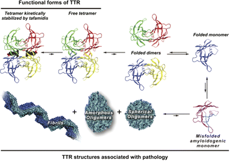
 Transtyretiini (TTR) on tetrameerinen  molekyyli, jossa on runsasti  beetatuppirakenteita.
 https://link.springer.com/article/10.1007/s40120-015-0031-3
Jos   geenissä on mutaatioita, proteiinin laskostumiset  haittaantuvat  ( oikein laskostunut  monomeeri, oligomeeri ja  tetrameeri  vaaditaan normaalifunktioon).  TTR syntetisoituu maksassa; on   hoitona ollut maksansiirtokin, jotta saataisiin  normaalia TTR- molekyyliä muodostumaan.
Nyt on lödyetty lääkeaine, joka pystyy stabiloimaan  TTR tetrameeria funktionaaliseksi.  Tästä kerrotaan vuoden 2015 artikkelissa , jonka saa ylläolevasta linkistä.  Päivän lääkärilehti nyt 2018  antaa hyvät  uutiset  lääkkeen kliinisestä tehosta  kardiologian alueelta. Nimittäin  mutatoitunut TTR- proteiini  vikuuttaa sydäntäkin ja seurauksena ajan mittaan on kardiomyopatiaa.  Mutaatio on  hyvin rajoittunut, kyse vain muutaman   funktionaalisesti  olennaisen  aminohapon vaihtumisesta. Peptidien  kyky  laskostua  oikealla  tavalla  on rajoittunut.  Yleensää väärin laskostuneet proteiinit  hajoitetaan solukoneistossa, joka tarkkaa proteiinin laatua.  Normaalillakin transtyretiinillä on vain 1.9  vuorokauden elinikä plasmassa, saati sitten  laskostumattomalla. Tämä uusi lääke stabiloi tetrameerirakennetta, joten  transtyretiinille tulee  funktionaalista  aikaa, mikä lopulta  heijastuu elinajan pidentymisessä ja oireitten   vähentymisessä  eri elimistä. Mm  hermoston taholta on havaittu kävelykyvyn kohenevan.
Tämä TTR- proteiini ja geeni kuuluu  eräänlaiseen chromeroniin, johon kuuluu monta hyvin olennaista  geeniä kattaen  usean funktion (A-vitamiinin toiminta-alue, kilpirauhasen toiminta -alue, jopa  sokeriaineenvaihdunta,  beetasolun funktio), joten  yksin  TTR- proteiinin funktion  kohentaminen  ei kohota potilaiden elämänlaatua, ellei  koko chromeronin  perusfunktiot koeteta myös pitää referensseissä.  Tuollaisia yhden mutaation tauteja  voidaan ehkä tulevaisuudessa vielä tehokkaamminkin hoitaa.
Tämä  tafamidis on hyvä uutinen.
.

 Fig. 1