Leta i den här bloggen

tisdag 21 augusti 2018

Lähdekirjallisuus: Fosfodiesteraaseista ja Fosfodiesteraasi-estäjistä

https://en.wikipedia.org/wiki/Phosphodiesterase_inhibitor
Tässä lähettssä on ei-selektiivisistä ja selektiivisitä PDE-estäjistä luetteloa. 


Fosfodiesteraasit PDE-perhe on entsyymejä, jotka  katkaisevat fosfodiesterisillan. Esim  sykliset fiosfodiesteraasit ovat  metaboloimassa cAMP ja cGMP siten,  että  kahdella esterisidoksella liittynyt sykliseksi muuttunut AMP palaa takaisin AMP- muotoon.
 Siis jos estetään PDE  funktiota,  säilyy cAMP ja cGMP signaali kauemman aikaa.

 PDE perheessa on   1,2,3, 10, ja 11 jotka  irrottavat  fosfodiesterisiltaa molemmista   sekä cAMP että cGMP molekyylistä, siis oikaisevat AMP:n
PDE 4, 7 ja 8 katkaisevat  esterisillat  cAMP:stä
 PDE 5,6, ja 9 katkaisevat   cGMP: pstä
 Nämä lienevät  kudostyypillisiä entsyymejä

Aivoissa   lähinnä PDE1B ja PDE4  esiintyvät  frontaalikorteksissa,
 PDE4, jokin muu  PDE? ja  PDE10A striatumissa . käsitelty  artikkelissa. edellä.
 Tämä PDE entsyymien vaikuttama  modulointi tapahtuu  kortikostriaalisissa  neuronipiireissä, joka näyttää olevan se  ongelmakohta  useissa neuropsykiatrisisisa  ja  neurologisissa  tiloissa. 


Muistiin alustavasti  21.8.2018 Tässä lähdeartikkelissa on paljon luettavaa vielä!  Se on tältä vuodelta 2018.
 https://www.sciencedirect.com/science/article/pii/S0149763417306450?via%3Dihub

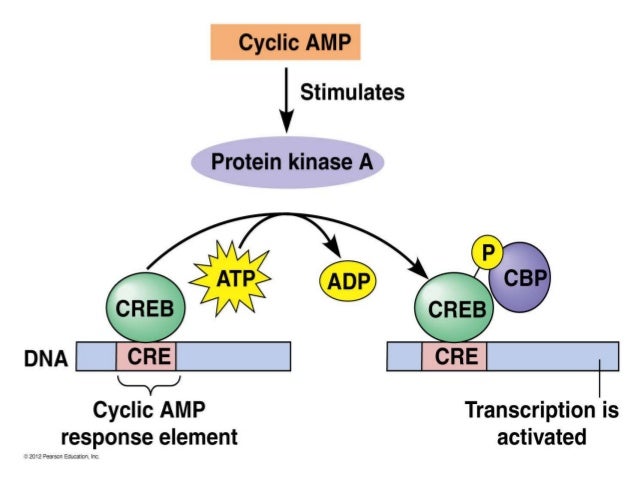
Tääsä huomaan että cAMP muuttuminen AMP muotoon tuottaa edelleen  toisen  vaikuttavan molekyylin. 
Miksi sen syklinen muoto on olennaista ?
Syklinen muoto omaa  geenisäätelyssä vaste-elementin.
https://image.slidesharecdn.com/slidesharesecondmessengersaj-140620103336-phpapp01/95/slideshare-second-messengers-aj-19-638.jpg?cb=1489738838
 https://image.slidesharecdn.com/slidesharesecondmessengersaj-140620103336-phpapp01/95/slideshare-second-messengers-aj-19-638.jpg?cb=1489738838


måndag 20 augusti 2018

Guanylyylisyklaasista 15 lähdettä, geenit

 Huomaan että tämä cGMP muodostus on  hyvin altis epätasapainolle ja  guanylyylisyklaasialueella on  kaikenaisia tauteja. En löytänyt tästä nyt erikseen NO-GC1 tai NO-GC2  nimisena neuronin  transmityeritasapainoon vaikuttavia  geenejä mainittuna  juuri näillä nimillä, joten  en tiedä mihin kromosomiin ne projisoituvat ja  mihin tauteihin esim neuroendokrinologisiin ne assosioituisivat. Jätän tähän ja jos tulee vastaan,  lisään  tästä  GLU-GABA- neuronaalisen   vapautumisen  tasapainon hienosäädöstä, niin laitan  sitten myöhemmin. Sekin tasapaino  ilmeisesti  on  mennyt ihmiskunnassa  vinoon, siten että normaalitasapainoa ei  enää saada takaisin ainakaan ilman strategioiden muuttamista.  GLU-GABA tasapainoa voisi sanoa tavallaan "neuronin kävelytahdiksi" sikäli että kävellessä täytyy todella olla  järjestys saman asian hyväksi, tietysti luonnossa kyllä voi mennä kuin tintti, mutta tässä vertauskuvallisesti: toinen jalka  alkaa yhteen suuntaan ja automaattisesti toinen jatkaa saman suuntaan asian hyväksi"   tämä on maksimaalisesti  näkyvissä niissä  häiriöissä, mitkä tulee motoriikassa  PD-taudissa, mutta  häiriö voi alkaa jo tästä GLU-GABA alkuvapautumisin  minimaalisen  aikaeron  hienosäädön katoamisesta.  Joka ainut tahdonalainen impulssi  menee hierarkisen hienosäätöverkoston modulatioon.


Nimiä
CUCY1A1 = CUCY1A3 = CUCA3  Kr.4    
 Soluble guanylate cyclases are heterodimeric proteins that catalyze the conversion of GTP to 3',5'-cyclic GMP and pyrophosphate. The protein encoded by this gene is an alpha subunit of this complex and it interacts with a beta subunit to form the guanylate cyclase enzyme, which is activated by nitric oxide, NO. Several transcript variants encoding a few different isoforms have been found for this gene. [provided by RefSeq, Jan 2012]
CUCY1A2, Kr.11 
 Soluble guanylate cyclases are heterodimeric proteins that catalyze the conversion of GTP to 3',5'-cyclic GMP and pyrophosphate. The protein encoded by this gene is an alpha subunit of this complex and it interacts with a beta subunit to form the guanylate cyclase enzyme, which is activated by nitric oxide,NO.  Two transcript variants encoding different isoforms have been found for this gene. [provided by RefSeq, Jan 2012]Expression Broad expression in endometrium (RPKM 8.8), placenta (RPKM 7.4) and 15 other tissues

GUCY1A4 =CUSY2D  Kr.17,  Retinal 
This gene encodes a retina-specific guanylate cyclase, which is a member of the membrane guanylyl cyclase family. Like other membrane guanylyl cyclases, this enzyme has a hydrophobic amino-terminal signal sequence followed by a large extracellular domain, a single membrane spanning domain, a kinase homology domain, and a guanylyl cyclase catalytic domain. In contrast to other membrane guanylyl cyclases, this enzyme is not activated by natriuretic peptides. Mutations in this gene result in Leber congenital amaurosis and cone-rod dystrophy-6 diseases.

NPR1 = CUCY2A , Kr.1
 This gene encodes natriuretic peptide receptor B, one of two integral membrane receptors for natriuretic peptides. Both NPR1 and NPR2 contain five functional domains: an extracellular ligand-binding domain, a single membrane-spanning region, and intracellularly a protein kinase homology domain, a helical hinge region involved in oligomerization, and a carboxyl-terminal guanylyl cyclase catalytic domain. The protein is the primary receptor for C-type natriuretic peptide (CNP), which upon ligand binding exhibits greatly increased guanylyl cyclase activity. Mutations in this gene are the cause of acromesomelic dysplasia Maroteaux type. [provided by RefSeq, Jul 2008]
 NPR2= CUCY2B, Kr,.9
Guanylyl cyclases, catalyzing the production of cGMP from GTP, are classified as soluble and membrane forms (Garbers and Lowe, 1994 [PubMed 7982997]). The membrane guanylyl cyclases, often termed guanylyl cyclases A through F, form a family of cell-surface receptors with a similar topographic structure: an extracellular ligand-binding domain, a single membrane-spanning domain, and an intracellular region that contains a protein kinase-like domain and a cyclase catalytic domain. GC-A and GC-B function as receptors for natriuretic peptides; they are also referred to as atrial natriuretic peptide receptor A (NPR1) and type B (NPR2; MIM 108961). Also see NPR3 (MIM 108962), which encodes a protein with only the ligand-binding transmembrane and 37-amino acid cytoplasmic domains. NPR1 is a membrane-bound guanylate cyclase that serves as the receptor for both atrial and brain natriuretic peptides (ANP (MIM 108780) and BNP (MIM 600295), respectively).[supplied by OMIM, May 2009]

GUCY2C Kr.12 
 Guanylate cyclase 2C
This observational study demonstrated the protein expression of GCC across various gastrointestinal malignancies. In all cancer histotypes, GCC protein localization was observed predominantly in the cytoplasm compared to the membrane region of tumor cells. Consistent immunohistochemistry detection of GCC protein expression in primary colorectal cancers and in their matched liver metastases suggests that the expression of GCC is maintained throughout the process of tumor progression and formation of metastatic disease.
CUSY2F , Kr.X 
The protein encoded by this gene is a guanylyl cyclase found predominantly in photoreceptors in the retina. The encoded protein is thought to be involved in resynthesis of cGMP after light activation of the visual signal transduction cascade, allowing a return to the dark state. This protein is a single-pass type I membrane protein. Defects in this gene may be a cause of X-linked retinitis pigmentosa. [provided by RefSeq, Dec 2008]
CUCA 1B  =CUCA2 , Kr.6


Search results

Items: 15

  • Showing Current items.
Name/Gene IDDescriptionLocationAliasesMIM
ID: 2984
guanylate cyclase 2C [Homo sapiens (human)]Chromosome 12, NC_000012.12 (14612632..14696625, complement)DIAR6, GC-C, GUC2C, MECIL, MUCIL, STAR601330
ID: 4881
natriuretic peptide receptor 1 [Homo sapiens (human)]Chromosome 1, NC_000001.11 (153678649..153693992)ANPRA, ANPa, GUC2A, GUCY2A, NPRA108960
ID: 3000
guanylate cyclase 2D, retinal [Homo sapiens (human)]Chromosome 17, NC_000017.11 (8002670..8020340)CACD1, CORD5, CORD6, CYGD, GUC1A4, GUC2D, LCA, LCA1, RCD2, RETGC-1, ROS-GC1, ROSGC, retGC600179
ID: 4882
natriuretic peptide receptor 2 [Homo sapiens (human)]Chromosome 9, NC_000009.12 (35782086..35809731)AMDM, ANPRB, ANPb, ECDM, GUC2B, GUCY2B, NPRB, NPRBi, SNSK108961
ID: 2641
glucagon [Homo sapiens (human)]Chromosome 2, NC_000002.12 (162142869..162152404, complement)GLP-1, GLP1, GLP2, GRPP138030
ID: 111
adenylate cyclase 5 [Homo sapiens (human)]Chromosome 3, NC_000003.12 (123282296..123448988, complement)AC5, FDFM600293
ID: 2982
guanylate cyclase 1 soluble subunit alpha 1 [Homo sapiens (human)]Chromosome 4, NC_000004.12 (155666710..155737062)GC-SA3, GUC1A3, GUCA3, GUCSA3, GUCY1A3, MYMY6139396
ID: 107
adenylate cyclase 1 [Homo sapiens (human)]Chromosome 7, NC_000007.14 (45574140..45723116)AC1, DFNB44103072
ID: 112
adenylate cyclase 6 [Homo sapiens (human)]Chromosome 12, NC_000012.12 (48766191..48789096, complement)AC6, LCCS8600294
ID: 114
adenylate cyclase 8 [Homo sapiens (human)]Chromosome 8, NC_000008.11 (130780300..131041604, complement)AC8, ADCY3, HBAC1103070
ID: 2986
guanylate cyclase 2F, retinal [Homo sapiens (human)]Chromosome X, NC_000023.11 (109372061..109482056, complement)CYGF, GC-F, GUC2DL, GUC2F, RETGC-2, ROS-GC2300041
ID: 2977
guanylate cyclase 1 soluble subunit alpha 2 [Homo sapiens (human)]Chromosome 11, NC_000011.10 (106674012..107018445, complement)GC-SA2, GUC1A2601244
ID: 108
adenylate cyclase 2 [Homo sapiens (human)]Chromosome 5, NC_000005.10 (7396230..7830081)AC2, HBAC2103071
ID: 196883
adenylate cyclase 4 [Homo sapiens (human)]Chromosome 14, NC_000014.9 (24318349..24335071, complement)AC4600292
ID: 2979
guanylate cyclase activator 1B [Homo sapiens (human)]Chromosome 6, NC_000006.12 (42183284..42194956, complement)GCAP2, GUCA2, RP48602275

Suoliston guanylyylisyklaasi ja suoliston kansantaudit IBS, Chron

https://www.ncbi.nlm.nih.gov/pubmed/28902124

Inflamm Bowel Dis. 2017 Oct;23(10):1752-1761. doi: 10.1097/MIB.0000000000001264.

Guanylate Cyclase C Activation Shapes the Intestinal Microbiota in Patients with Familial Diarrhea and Increased Susceptibility for Crohn's Disease.

Abstract

BACKGROUND:

With 25% prevalence of Crohn's disease, Familial GUCY2C diarrhea syndrome (FGDS) is a monogenic disorder potentially suited to study initiating factors in inflammatory bowel disease (IBD). We aimed to characterize the impact of an activating GUCY2C mutation on the gut microbiota in patients with FGDS controlling for Crohn's disease status and to determine whether changes share features with those observed in unrelated patients with IBD.

METHODS:

Bacterial DNA from fecal samples collected from patients with FGDS (N = 20), healthy relatives (N = 11), unrelated healthy individuals (N = 263), and IBD controls (N = 46) was subjected to sequencing of the V3-V4 region of the 16S rRNA gene to determine gut microbiota composition. Food frequency questionnaires were obtained from patients with FGDS and their relatives.

RESULTS:

Compared with healthy controls, FGDS displayed prominent changes in many microbial lineages including increase in Enterobacteriaceae, loss of Bifidobacterium and Faecalibacterium prausnitzii but an unchanged intraindividual (alpha) diversity. The depletion of F. prausnitzii is in line with what is typically observed in Crohn's disease. There was no significant difference in the dietary profile between the patients and related controls. The gut microbiota in related and unrelated healthy controls was also similar, suggesting that diet and familial factors do not explain the gut microbiota alterations in FGDS.

CONCLUSIONS:

The findings support that the activating mutation in GUCY2C creates an intestinal environment with a major influence on the microbiota, which could contribute to the increased susceptibility to IBD in patients with FGDS.
PMID:
28902124
DOI:
10.1097/MIB.0000000000001264
[Indexed for MEDLINE]

Sydämen sähköinen toiminta, NO-GC1-cGMP -tie sydämen homeostaasi

https://www.ncbi.nlm.nih.gov/pubmed/29269353
J Am Heart Assoc. 2017 Dec 21;6(12). pii: e006397. doi: 10.1161/JAHA.117.006397.

Newly Identified NO-Sensor Guanylyl Cyclase/Connexin 43 Association Is Involved in Cardiac Electrical Function.

Abstract

BACKGROUND:

Guanylyl cyclase, a heme-containing α1β1 heterodimer (GC1), produces cGMP in response to Nitric oxide (NO) stimulation. The NO-GC1-cGMP pathway negatively regulates cardiomyocyte contractility and protects against cardiac hypertrophy-related remodeling. We recently reported that the β1 subunit of GC1 is detected at the intercalated disc with connexin 43 (Cx43). Cx43 forms gap junctions (GJs) at the intercalated disc that are responsible for electrical propagation. We sought to determine whether there is a functional association between GC1 and Cx43 and its role in cardiac homeostasis.

METHODS AND RESULTS:

GC1 and Cx43 immunostaining at the intercalated disc and coimmunoprecipitation from membrane fraction indicate that GC1 and Cx43 are associated. Mice lacking the α subunit of GC1 (GCα1 knockout mice) displayed a significant decrease in GJ function (dye-spread assay) and Cx43 membrane lateralization. In a cardiac-hypertrophic model, angiotensin II treatment disrupted the GC1-Cx43 association and induced significant Cx43 membrane lateralization, which was exacerbated in GCα1 knockout mice. Cx43 lateralization correlated with decreased Cx43-containing GJs at the intercalated disc, predictors of electrical dysfunction. Accordingly, an ECG revealed that angiotensin II-treated GCα1 knockout mice had impaired ventricular electrical propagation. The phosphorylation level of Cx43 at serine 365, a protein-kinase A upregulated site involved in trafficking/assembly of GJs, was decreased in these models.

CONCLUSIONS:

GC1 modulates ventricular Cx43 location, hence GJ function, and partially protects from electrical dysfunction in an angiotensin II hypertrophy model. Disruption of the NO-cGMP pathway is associated with cardiac electrical disturbance and abnormal Cx43 phosphorylation. This previously unknown NO/Cx43 signaling could be a protective mechanism against stress-induced arrhythmia.

KEYWORDS:

Nitric oxide; arrhythmia; cGMP; cardiac function; cardiovascular disease; connexin 43; guanylyl cyclase
PMID:
29269353
PMCID:
PMC5778997
DOI:
10.1161/JAHA.117.006397

Aivan uutta jatkoa näistä cGMP -muodostavista neuronaalisista balansoivista entsyymeistä (hiiren aivossa)

https://www.ncbi.nlm.nih.gov/pubmed/30087260
Suom. Huonontunut NO-cGMP-signalointi on liittynyt useisiin neurologisiin tauteihin.  NO-sensitiiviset guanylyylisyklaasit (NO-GC) , joista kaksi isoformia tunnetaan, edustavat  lupaava lääkekohdetta aivojen cGMP-pitoisuuden  kohottamiseksi. 
  On jo löydetty joitain pieniä lääkkeen kaltaisia molekyylejä, jotka vaikuttavat synergistisesti NO:n kanssa  stimuloiden NO-GC entsyymiaktiivisututa. Kuitenkaan näiden stimulaatorien aivovaikutuksia ei vielä oiken ymmärretä  ja tämä artikkeli   kertoo  yksityiskohtaisista tutkimuksista  hiirellä ja johtopäätös on: On tullut selväksi jo, että NO-GC- entsyymistimulaattorit voivat lisätä cGMP-signalointia neuronissa ja niitä pitäisi kehittää  edelleen neurologisten tautien hoitoon.
Int J Mol Sci. 2018 Aug 7;19(8). pii: E2313. doi: 10.3390/ijms19082313.
cGMP Imaging in Brain Slices Reveals Brain Region-Specific Activity of NO-Sensitive Guanylyl Cyclases (NO-GCs) and NO-GC Stimulators.

Abstract

Impaired NO-cGMP signaling has been linked to several neurological disorders. NO-sensitive guanylyl cyclase (NO-GC), of which two isoforms-NO-GC1 and NO-GC2-are known, represents a promising drug target to increase cGMP in the brain. Drug-like small molecules have been discovered that work synergistically with NO to stimulate NO-GC activity. However, the effects of NO-GC stimulators in the brain are not well understood.

 In the present study, we used Förster/fluorescence resonance energy transfer (FRET)-based real-time imaging of cGMP in acute brain slices and primary neurons of cGMP sensor mice to comparatively assess the activity of two structurally different NO-GC stimulators, IWP-051 and BAY 41-2272, in the cerebellum, striatum and hippocampus. BAY 41-2272 potentiated an elevation of cGMP induced by the NO donor DEA/NO in all tested brain regions. Interestingly, IWP-051 potentiated DEA/NO-induced cGMP increases in the cerebellum and striatum, but not in the hippocampal CA1 area or primary hippocampal neurons. The brain-region-selective activity of IWP-051 suggested that it might act in a NO-GC isoform-selective manner.

Results of mRNA in situ hybridization indicated that the cerebellum and striatum express NO-GC1 and NO-GC2, while the hippocampal CA1 area expresses mainly NO-GC2.
  IWP-051-potentiated DEA/NO-induced cGMP signals in the striatum of NO-GC2 knockout mice but was ineffective in the striatum of NO-GC1 knockout mice. These results indicate that IWP-051 preferentially stimulates NO-GC1 signaling in brain slices.

Interestingly, no evidence for an isoform-specific effect of IWP-051 was observed when the cGMP-forming activity of whole brain homogenates was measured. This apparent discrepancy suggests that the method and conditions of cGMP measurement can influence results with NO-GC stimulators. Nevertheless, it is clear that NO-GC stimulators enhance cGMP signaling in the brain and should be further developed for the treatment of neurological diseases.

KEYWORDS:

Cyclic GMP; FRET imaging; NO-GC stimulators; Purkinje cells; cerebellar granule cells; guanylyl cyclase; hippocampal neurons; nitric oxide; striatum; transgenic mice
PMID:
30087260
DOI:
10.3390/ijms19082313

Neuronin GLU ja GABA tasapainon hienosäädöstä cGMP:n avulla

https://www.ncbi.nlm.nih.gov/pubmed/28782641
Tässä on  hypoglutamaattiteoriasta  myös, mutta tässä nähdään neuronaalisen  glutamaatin vähäisen  pitoisuuden taustasyyksi   toisen   excitatorisen  aminohapon  entsyymin  DDO stimuloivan vaikutuksen puute  glutamaatin muodostukseen  ja tästä teoria saa laajentumista .

Sci Rep. 2017; 7: 46288.
Published online 2017 Apr 10. doi:  10.1038/srep46288
PMCID: PMC5385520
PMID: 28393897
Olanzapine, but not clozapine, increases glutamate release in the prefrontal cortex of freely moving mice by inhibiting D-aspartate oxidase activitySilvia Sacchi,1,2,* Vito De Novellis et al. 

Kaksi guanyylisyklaasia osallistuu  NO (typpioksidin) -vaikutusten johtamiseen synaptisessa  transmissiossa hippokampialueessa.  Entsyymit ovat NO-GC1 ja  NOGC2. Tutkijat halusivat  selvittää entsyymi- isoformien  osuuden muissa  aivoalueissa. He käyttivät ensinmainitun entsyymin suhteen poistogeenista hiirtä ja analysoivat sitten  somatosensorisesta kuorikerroksesta GLU-ergisen ja GABA-ergisen  hermovälityksen kerroksen -II/III pyramidaalisista neuroneista. Poistogeenisellä hiirellä, jolta  ensimmäinen  entsyymi  (NO-GC1) puuttui, ilmeni alentuneita  excitatorisia ja inhibitorisia  postsynaptisia  miniatyyrivirtauksia, kohonnutta paired-pulse  tahtia ja alentunutta  input-output- käyrää herätesignaaleista- mikä viittaa siihen, että  sekä  GLU että  GABA-vapautumiset olivat alentuneet tässä  poistogeenisessa hiiressä.  Nämä funktioanaliset muutokset  poistogeenisessä hiiressä  johtuivat  syklisen GMP:n puutteesta, koska  sen analogilla saatettiin palauttaa  aminohappojen vapautumiset  ennalleen kuten   wt-hiiressä. 
Etsittäessä  cGMP kohteita  normaalihiirella(wt) kaksi HCN-kanavan blokkaria  vähensi glutamaatin vaaputumsita samalle tasolle kuin mainitussa poistogeenisessa  tapauksessa . Tämä viittaisi näiden  HNC-kanavien olevan  mahdollisia presynaoptisesti mudosotuneen cGMP:n effektoreita , jotka  lisäävät  glutamaatin vaaputumisen mahdollisuutta. HCN kanavat ovat  hyperpolarisaation aktivoimia  syklisten nukleotidiensäätelemiä kanavia. 
Entä sitten jos blokeerattiin postsynaptinen GLU -reseptori NMDA? Siinä tapauksessa   NMDA-reseptorista riippuvainen NO-signaali  näytti  linkkiytyvän  NO-GC1 -entsyymin vaikuttamaan  presynaptisen GABA:n  vapautumiseen.  On huomattava, että tasapaino GLU-ergisten ja GABA-ergisten input-(sisään)syöttöjen  kesken  yksittäisissä synapseissa pysyi muuttumattomana  poistogeenisessa hiiressä. Yhteenvetona tulokset viittaavat siihen, että  presynaptisen entsyymin NO-GC1 generoima syklinen GMP ( cGMP) omaa tehtävän hienosäätää inhibitoriset ja excitatoriset  input (sisään)syötöt  somatosensorisen harmaan aivokuoren yksittäisissä  synapseissa.

Abstract

In hippocampus, two guanylyl cyclases (NO-GC1 and NO-GC2) are involved in the transduction of the effects of nitric oxide (NO) on synaptic transmission. However, the respective roles of the NO-GC isoforms on synaptic transmission are less clear in other regions of the brain. In the present study, we used knock-out mice deficient for the NO-GC1 isoform (NO-GC1 KO) to analyze its role in the glutamatergic and GABAergic neurotransmission at pyramidal neurons in layers II/III of somatosensory cortex. NO-GC1 KO slices revealed reduced frequencies of miniature excitatory- and inhibitory-postsynaptic currents, increased paired-pulse ratios and decreased input-output curves of evoked signals, which indicated the reduction of glutamate and GABA release in NO-GC1 KO mice. The functional changes in NO-GC1 KO mice were caused by the lack of cGMP as they were rescued to WT-like levels by the cGMP analog, 8-Br-PET-cGMP and conversely, mimicked by the NO-GC inhibitor, ODQ, in WT slices.
 In search of a cGMP target, two blockers of hyperpolarization-activated cyclic nucleotide-gated (HCN) channels (ZD7288 and DK-AH269) reduced glutamate release in WT to the level of NO-GC1 KO mice suggesting HCN channels as possible effectors for presynaptic cGMP enhancing the glutamate release probability.
By blocking postsynaptic NMDA receptors, the NMDA receptor-dependent NO signal was shown to be linked to the effect of NO-GC1 on presynaptic GABA release. Of note, the balance between glutamatergic and GABAergic inputs at individual synapses remained unaltered in the NO-GC1 KO mice. In sum, our results indicate a role for cGMP generated by presynaptic localized NO-GC1 to adjust inhibitory and excitatory inputs at individual synapses in the somatosensory cortex.

KEYWORDS:

GABA; NO-GC1; cGMP; glutamate; somatosensory cortex

tisdag 10 juli 2018

Vahvin excitotoksiini hermostossa on kinolinaatti

kinolinaatti (quinolinate) kuuluu tryptofaanin metaboliseen akrttaan. tryptofaani on  vain ravinnosta saatava aminohappo. Luonnollsiesti katabolia voi vapauttaa kehossa  tryptofaaneja rakenteista.
Normaalisti kinolinaatti  voi dekarboksyloitua tärkeäksi nikotiinihapoksi, josta keho valmistaa koentsyymejä NAD+, NADP .
olin tässä katsomassa NAD+  syntyteitä, lukiessani fundamentaalista sirtuiini-etikkahappo  pistettä  solussa.
Siinä muodostuu  syklisesi  PRPP  phosphoribosyylipyrofosfaattia joka sitten uudestaan rakentuu NAD+ molekyyliin. 
Forsforibosyylitransferaaseja on 3 eri entsyymiä. Yksi  (NaPRT) käyttää valmista  nikotiinihappoa  (Na)  jota on dietääristi muodostunut  ja toinen  käyttää nikotiinihappoamidimuotoa NAM,  (NAMPRT)  ja yksi käyttää kinolinaattimuotoa. QPRT, (QPRTase).
Koska tästä kinolinaatti- kohdasta on pahaa mainetta aivotoksisena pisteenä, haen  geenistä QPRT jonkin  sitaatin.  Epileptisessä aivossa  tämän entsyymin pitoisuus on alentunut.  Vaurioituneessa aivossa myös, koska tryptofaaniaineenvaihdunnan  kynureenitie aktivoituu ja kinolinaatin tuotanto nousee.

(Huom:  tässä saattaisi olla merkitystä huomioida  eksogeenisen tryptofaanin  dietääriset pitoisuudet  ja myös  valmiin  päätevitamiinin   mahdollinen  merkitys  metabolialle sekä  aineenvaihduntareitillä välttämättömien  tekij'kirjon     saanti.  B6-vitamiini on erityisesti  kynureenimetaboliassa    kinolinaatin (quinolinate)   nikotiinihapoksi muuttava  tekijä. On sanottu, että 60 mg tryptofaania tuotaa  1 mg "omaa B-vitamiinia" nikotiinihappoa- tietysti  entsyymien toimiessa-  Entä jos on jokin vaurio joka tuottaa  nikotiinihappoa edeltävää kinolinaattia liikaa?  tai  QPRT entsyymi on heikentynyt? - toksinen tekijä siaitsee silloin  tryptofaania aineenvaihduntaa  suorittavissa  soluissa.

Kynureniinini (KYN) voi tuottaa  tuottaa KYNA , neuroprotektiivista  molekyyliä Kynurenic acid.Se on komplementaarinen antagonisoi  NMDAR reseptorin Gly-kohdalle.
B6 vitamiinin puutteesa KYN  muuttuu xantureenihapoksi .
KYN oksidaatio johtaa 3-hyroksykunureeniin ja  siitä käsin tulee välivaiheita myöten  myös kinolinaatti (Quinolinate) ja normaalein entsyymein nikotiinihappoa.
Tämä viimeksi mainittu tie  kynureniinista  (stressituote)  eteenpäin  aktivoituu vauriosta.

 https://www.ncbi.nlm.nih.gov/gene/23475

QPRT geeni (16p11.2)
Also known as QPRTase; HEL-S-90n
Summary This gene encodes a key enzyme in catabolism of quinolinate, an intermediate in the tryptophan-nicotinamide adenine dinucleotide  (NAD) pathway. Quinolinate acts as a most potent endogenous exitotoxin to neurons. Elevation of quinolinate levels in the brain has been linked to the pathogenesis of neurodegenerative disorders such as epilepsy, Alzheimer's disease, and Huntington's disease. Alternative splicing results in multiple transcript variants. [provided by RefSeq, Dec 2015]
 

GeneRIFs: Gene References Into FunctionsWhat's a GeneRIF?

 https://www.researchgate.net/profile/Simon_Jones21/publication/257350797/figure/fig1/AS:601612707979264@1520447055044/Overview-of-the-kynurenine-pathway-of-tryptophan-metabolism-note-Key-enzymes-are.png
Overview of the kynurenine pathway of tryptophan metabolism. note: Key enzymes are indicated in red. Abbreviations: IDO, Indoleamine 2,3-dioxygenase; TDO2, Tryptophan 2,3-dioxygenase; KYNU, Kynureninase; KATs, Kynurenine aminotransferases; KMO, Kynurenine 3-monooxygenase; 3HAO, 3-hydroxyanthranilic acid oxygenase; ACMSD, Aminocarboxymuconate-semialdehyde decarboxylase; QPRT, quinolinic acid phosphoribosyltransferase.